- Case Report
- Open access
- Published: 07 August 2009

Primary abdominal ectopic pregnancy: a case report
- Recep Yildizhan 1 ,
- Ali Kolusari 1 ,
- Fulya Adali 2 ,
- Ertan Adali 1 ,
- Mertihan Kurdoglu 1 ,
- Cagdas Ozgokce 1 &
- Numan Cim 1
Cases Journal volume 2 , Article number: 8485 ( 2009 ) Cite this article
21k Accesses
18 Citations
Metrics details
Introduction
We present a case of a 13-week abdominal pregnancy evaluated with ultrasound and magnetic resonance imaging.
Case presentation
A 34-year-old woman, (gravida 2, para 1) suffering from lower abdominal pain and slight vaginal bleeding was transferred to our hospital. A transabdominal ultrasound and magnetic resonance imaging were performed. The diagnosis of primary abdominal pregnancy was confirmed according to Studdiford's criteria. A laparatomy was carried out. The placenta was attached to the mesentery of sigmoid colon and to the left abdominal sidewall. The placenta was dissected away completely and safely. No postoperative complications were observed.
Ultrasound examination is the usual diagnostic procedure of choice. In addition magnetic resonance imaging can be useful to show the localization of the placenta preoperatively.
Abdominal pregnancy, with a diagnosis of one per 10000 births, is an extremely rare and serious form of extrauterine gestation [ 1 ]. Abdominal pregnancies account for almost 1% of ectopic pregnancies [ 2 ]. It has reported incidence of one in 2200 to one in 10,200 of all pregnancies [ 3 ]. The gestational sac is implanted outside the uterus, ovaries, and fallopian tubes. The maternal mortality rate can be as high as 20% [ 3 ]. This is primarily because of the risk of massive hemorrhage from partial or total placental separation. The placenta can be attached to the uterine wall, bowel, mesentery, liver, spleen, bladder and ligaments. It can be detach at any time during pregnancy leading to torrential blood loss [ 4 ]. Accurate localization of the placenta pre-operatively could minimize blood loss during surgery by avoiding incision into the placenta [ 5 ]. It is thought that abdominal pregnancy is more common in developing countries, probably because of the high frequency of pelvic inflammatory disease in these areas [ 6 ]. Abdominal pregnancy is classified as primary or secondary. The diagnosis of primary abdominal pregnancy was confirmed according to Studdiford's criteria [ 7 ]. In these criteria, the diagnosis of primary abdominal pregnancy is based on the following anatomic conditions: 1) normal tubes and ovaries, 2) absence of an uteroplacental fistula, and 3) attachment exclusively to a peritoneal surface early enough in gestation to eliminate the likelihood of secondary implantation. The placenta sits on the intra-abdominal organs generally the bowel or mesentery, or the peritoneum, and has sufficient blood supply. Sonography is considered the front-line diagnostic imaging method, with magnetic resonance imaging (MRI) serving as an adjunct in cases when sonography is equivocal and in cases when the delineation of anatomic relationships may alter the surgical approach [ 8 ]. We report the management of a primary abdominal pregnancy at 13 weeks.
The patient was a 34-year-old Turkish woman, gravida 2 para 1 with a normal vaginal delivery 15 years previously. Although she had not used any contraceptive method afterwards, she had not become pregnant. She was transferred to our hospital from her local clinic at the gestation stage of 13 weeks because of pain in the lower abdomen and slight vaginal bleeding. She did not know when her last menstrual period had been, due to irregular periods. At admission, she presented with a history of abdominal distention together with steadily increasing abdominal and back pain, weakness, lack of appetite, and restlessness with minimal vaginal bleeding. She denied a history of pelvic inflammatory disease, sexually transmitted disease, surgical operations, or allergies. Blood pressure and pulse rate were normal. Laboratory parameters were normal, with a hemoglobin concentration of 10.0 g/dl and hematocrit of 29.1%. Transvaginal ultrasonographic scanning revealed an empty uterus with an endometrium 15 mm thick. A transabdominal ultrasound (Figure 1 ) examination demonstrated an amount of free peritoneal fluid and the nonviable fetus at 13 weeks without a sac; the placenta measured 58 × 65 × 67 mm. Abdominal-Pelvic MRI (Philips Intera 1.5T, Philips Medical Systems, Andover, MA) in coronal, axial, and sagittal planes was performed especially for localization of the placenta before she underwent surgery. A non-contrast SPAIR sagittal T2-weighted MRI strongly suggested placental invasion of the sigmoid colon (Figure 2 ).
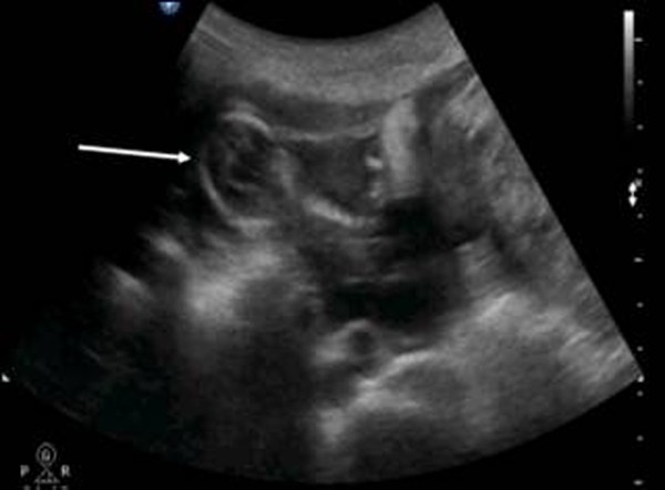
Pelvic ultrasound scanning . Diffuse free intraperitoneal fluid was seen around the fetus and small bowel loops.
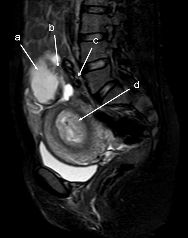
T2W SPAIR sagittal MRI of lower abdomen demonstrating the placental invasion . Placenta (a) , invasion area (b) , sigmoid colon (c) , uterine cavity (d) .
Under general anesthesia, a median laparotomy was performed and a moderate amount of intra-abdominal serohemorrhagic fluid was evident. The placenta was attached tightly to the mesentery of sigmoid colon and was loosely adhered to the left abdominal sidewall (Figure 3 ). The fetus was localized at the right of the abdomen and was related to the placenta by a chord. The placenta was dissected away completely and safely from the mesentery of sigmoid colon and the left abdominal sidewall. Left salpingectomy for unilateral hydrosalpinx was conducted. Both ovaries were conserved. After closure of the abdominal wall, dilatation and curettage were also performed but no trophoblastic tissue was found in the uterine cavity. As a management protocol in our department, we perform uterine curettage in all patients with ectopic pregnancy gently at the end of the operation, not only for the differential diagnosis of ectopic pregnancy, but also to help in reducing present or possible postoperative vaginal bleeding.
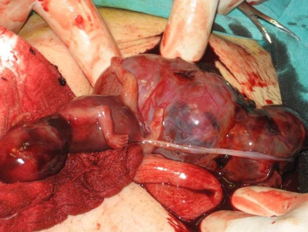
Fetus, placenta and bowels .
The patient was awakened, extubated, and sent to the room. The patient was discharged on post-operative day five with the standard of care at our hospital.
In the present case, we were able to demonstrate primary abdominal pregnancy according to Studdiford's criteria with the use of transvaginal and transabdominal ultrasound examination and MRI. In our case, both fallopian tubes and ovaries were intact. With regard to the second criterion, we did not observe any uteroplacental fistulae in our case. Since abdominal pregnancy at less than 20 weeks of gestation is considered early [ 9 ], our case can be regarded as early, and so we dismissed the possibility of secondary implantation.
The recent use of progesterone-only pills and intrauterine devices with a history of surgery, pelvic inflammatory disease, sexually transmitted disease, and allergy increases the risk of ectopic pregnancy. Our patient had not been using any contraception, and did not report a history of the other risk factors.
The clinical presentation of an abdominal pregnancy can differ from that of a tubal pregnancy. Although there may be great variability in symptoms, severe lower abdominal pain is one of the most consistent findings [ 10 ]. In a study of 12 patients reported by Hallatt and Grove [ 11 ], vaginal bleeding occurred in six patients.
Ultrasound examination is the usual diagnostic procedure of choice, but the findings are sometimes questionable. They are dependent on the examiner's experience and the quality of the ultrasound. Transvaginal ultrasound is superior to transabdominal ultrasound in the evaluation of ectopic pregnancy since it allows a better view of the adnexa and uterine cavity. MRI provided additional information for patients who needed precise diagnosing. After the diagnosis of abdominal pregnancy became definitive, it was essential to determine the localization of the placenta. Meanwhile, MRI may help in surgical planning by evaluating the extent of mesenteric and uterine involvement [ 12 ]. Non-contrast MRI using T 2 -weighted imaging is a sensitive, specific, and accurate method for evaluating ectopic pregnancy [ 13 ], and we used it in our case.
Removal of the placental tissue is less difficult in early pregnancy as it is likely to be smaller and less vascular. Laparoscopic removal of more advanced abdominal ectopic pregnancies, where the placenta is larger and more invasive, is different [ 14 ]. Laparoscopic treatment must be considered for early abdominal pregnancy [ 15 ].
Complete removal of the placenta should be done only when the blood supply can be identified and careful ligation performed [ 11 ]. If the placenta is not removed completely, it has been estimated that the remnant can remain functional for approximately 50 days after the operation, and total regression of placental function is usually complete within 4 months [ 16 ].
In conclusion, ultrasound scanning plus MRI can be useful to demonstrate the anatomic relationship between the placenta and invasion area in order to be prepared preoperatively for the possible massive blood loss.
Written informed consent was obtained from the patient for publication of this case report and accompanying images. A copy of the written consent is available for review by the Editor-in-chief of this journal.
Abbreviations
Magnetic Resonance Imaging
Spectral Presaturation Attenuated by Inversion Recovery.
Yildizhan R, Kurdoglu M, Kolusari A, Erten R: Primary omental pregnancy. Saudi Med J. 2008, 29: 606-609.
PubMed Google Scholar
Ludwig M, Kaisi M, Bauer O, Diedrich K: The forgotten child-a case of heterotopic, intra-abdominal and intrauterine pregnancy carried to term. Hum Reprod. 1999, 14: 1372-1374. 10.1093/humrep/14.5.1372.
Article CAS PubMed Google Scholar
Alto WA: Abdominal pregnancy. Am Fam Physician. 1990, 41: 209-214.
CAS PubMed Google Scholar
Ang LP, Tan AC, Yeo SH: Abdominal pregnancy: a case report and literature review. Singapore Med J. 2000, 41: 454-457.
Martin JN, Sessums JK, Martin RW, Pryor JA, Morrison JC: Abdominal pregnancy: current concepts of management. Obstet Gynecol. 1988, 71: 549-557.
Maas DA, Slabber CF: Diagnosis and treatment of advanced extra-uterine pregnancy. S Afr Med J. 1975, 49: 2007-2010.
Studdiford WE: Primary peritoneal pregnancy. Am J Obstet Gynecol. 1942, 44: 487-491.
Google Scholar
Wagner A, Burchardt A: MR imaging in advanced abdominal pregnancy. Acta Radiol. 1995, 36: 193-195. 10.3109/02841859509173377.
Gaither K: Abdominal pregnancy-an obstetrical enigma. South Med J. 2007, 100: 347-348.
Article PubMed Google Scholar
Onan MA, Turp AB, Saltik A, Akyurek N, Taskiran C, Himmetoglu O: Primary omental pregnancy: case report. Hum Reprod. 2005, 20: 807-809. 10.1093/humrep/deh683.
Hallatt JG, Grove JA: Abdominal pregnancy: a study of twenty-one consecutive cases. Am J Obstet Gynecol. 1985, 152: 444-449.
Malian V, Lee JH: MR imaging and MR angiography of an abdominal pregnancy with placental infarction. AJR Am J Roentgenol. 2001, 177: 1305-1306.
Yoshigi J, Yashiro N, Kinoshito T, O'uchi T, Kitagaki H: Diagnosis of ectopic pregnancy with MRI: efficacy of T2-weighted imaging. Magn Reson Med Sci. 2006, 5: 25-32. 10.2463/mrms.5.25.
Kwok A, Chia KKM, Ford R, Lam A: Laparoscopic management of a case of abdominal ectopic pregnancy. Aust N Z J Obstet Gynaecol. 2002, 42: 300-302. 10.1111/j.0004-8666.2002.300_1.x.
Pisarska MD, Casson PR, Moise KJ, Di Maio DJ, Buster JE, Carson SA: Heterotopic abdominal pregnancy treated at laparoscopy. Fertil Steril. 1998, 70: 159-160. 10.1016/S0015-0282(98)00104-6.
France JT, Jackson P: Maternal plasma and urinary hormone levels during and after a successful abdominal pregnancy. Br J Obstet Gynaecol. 1980, 87: 356-362.
Download references
Author information
Authors and affiliations.
Department of Obstetrics and Gynecology, School of Medicine, Yuzuncu Yil University, Van, Turkey
Recep Yildizhan, Ali Kolusari, Ertan Adali, Mertihan Kurdoglu, Cagdas Ozgokce & Numan Cim
Department of Radiology, Women and Child Hospital, Van, Turkey
Fulya Adali
You can also search for this author in PubMed Google Scholar
Corresponding author
Correspondence to Recep Yildizhan .
Additional information
Competing interests.
The authors declare that they have no competing interests.
Authors' contributions
All authors were involved in patient's care. RY, AK and FA analyzed and interpreted the patient data regarding the clinical and radiological findings of the patient and prepared the manuscript. EA, MK and CO edit and coordinated the manuscript. All authors read and approved the final manuscript.
Rights and permissions
Open Access This article is published under license to BioMed Central Ltd. This is an Open Access article is distributed under the terms of the Creative Commons Attribution License ( https://creativecommons.org/licenses/by/2.0 ), which permits unrestricted use, distribution, and reproduction in any medium, provided the original work is properly cited.
Reprints and permissions
About this article
Cite this article.
Yildizhan, R., Kolusari, A., Adali, F. et al. Primary abdominal ectopic pregnancy: a case report. Cases Journal 2 , 8485 (2009). https://doi.org/10.4076/1757-1626-2-8485
Download citation
Received : 12 January 2009
Accepted : 19 June 2009
Published : 07 August 2009
DOI : https://doi.org/10.4076/1757-1626-2-8485
Share this article
Anyone you share the following link with will be able to read this content:
Sorry, a shareable link is not currently available for this article.
Provided by the Springer Nature SharedIt content-sharing initiative
- Sigmoid Colon
- Ectopic Pregnancy
- Pelvic Inflammatory Disease
- Lower Abdominal Pain
- Transabdominal Ultrasound
Cases Journal
ISSN: 1757-1626

Understanding and Managing Ectopic Pregnancy
Jan 25, 2023
90 likes | 197 Views
This PowerPoint presentation provides an overview of the diagnosis, and management of ectopic pregnancies. It discusses the risk factors for ectopic pregnancy, common diagnostic tests, and various treatment options. It also outlines common complications and strategies for patient care. The presentation is designed to provide healthcare providers with the knowledge and tools needed to effectively care for patients with ectopic pregnancy.<br><br>Website: https://rajasuperspecialityclinic.com/team-member/dr-abarajda-v/
Share Presentation

Presentation Transcript
UNDERSTANDING AND MANAGING ECTOPIC PREGNANCY A Guide for Patients Raja Superspeciality Clinic
ECTOPIC PREGNANCY Definition Ectopic pregnancy occurs when a fertilized egg implants outside of the uterus, typically in the fallopian tube. This type of pregnancy cannot progress normally and can be life- threatening if not treated promptly.
CAUSES OF ECTOPIC PREGNANCY Damage to the fallopian tubes, such as from infection or previous surgery Hormonal imbalances Certain medical conditions, such as endometriosis
SYMPTOMS OF ECTOPIC PREGNANCY Vaginal bleeding or spotting Abdominal or pelvic pain Shoulder pain (caused by bleeding into the abdominal cavity)
DIAGNOSIS AND TREATMENT Ectopic pregnancy is typically diagnosed with a combination of blood tests, pelvic examination, and imaging tests such as ultrasonography. Treatment options include medication (such as methotrexate) or surgery (such as laparoscopy or laparotomy)
PREVENTION OF ECTOPIC PREGNANCY Preventing STIs and seeking prompt treatment can reduce the risk of fallopian tube damage Using birth control can also reduce the risk of ectopic pregnancy
Contact Raja Superspeciality Clinic today to schedule an appointment and receive the best care for Ectopic pregnancy. For more information, contact us www.rajasuperspecialityclinic.com 860 860 1590
- More by User

Suspected Ectopic Pregnancy
Suspected Ectopic Pregnancy Obstetrics & Gynecology vol. 107, No2 part 1, February, 2006 OBGY R2 BYUN JUNG MI Suspected Ectopic Pregnancy Abstract Incidence Risk factors Diagnostic Approach Therapeutic Approach Follow-up Abstract
1.24k views • 54 slides

ectopic pregnancy
INCIDENCE >1 in 100 pregnancies. . Recent evidence indicates that the incidence of ectopic pregnancy has been rising in many countries.USA-5 foldUK-2 foldFrance 15/1000 pregnanciesIndia-1in100 deliveriesRecurrence rate - 15% after 1st, 25% after 2 ectopics. 26/08/2011 05:53. Ectopic Pregnancy.
3k views • 44 slides

Ectopic Pregnancy Ch 25
Ectopic Pregnancy Ch 25. 2009-2010 Academic Year MSIII Ob/Gyn Clerkship Self-Directed Study. Case Presentation.
606 views • 11 slides

ECTOPIC PREGNANCY
Any pregnancy where the fertilized ovum gets implanted and develops in a site other than normal the uterine cavity.963 AD Albucasis first described ectopic pregnancy.1884 - Robert Lawson Tait of Birmingham performed the first successful operation for ectopic pregnancy.. INTRODUCTION. 1 in 100
984 views • 31 slides
ECTOPIC PREGNANCY. Dr. Madhavi Karki. Definition. Sites of Implantation:. Fig: Implantation Sites in Ectopic Pregnancy. ETIOLOGY. History – Taking . Clinical Features of Ectopic Pregnancy. Investigations : . Differential Diagnosis. Management of ectopic pregnancy : . ACUTE :
864 views • 12 slides

Ectopic Pregnancy
Ectopic Pregnancy. A’asem Zeidan Abu- Shtaya. Normally, . Normally, . Fertilization occurs in the lateral third of the fallopian tube. On average it takes the spermatozoa 1 hr to reach the ovum.
2.71k views • 62 slides

Ectopic Pregnancy. Xiaofang Yi, M.D. Hospital of OB/GYN, Fudan University Email: yi.annie [email protected] Mobile: 15026585241. Abbreviations. STD : sexually transmitted disease ART : assisted reproductive technique hCG : human chorionic gonadotropin TVS : transvaginal sonography
3.18k views • 68 slides

ECTOPIC PREGNANCY. is implantation of the fertilized ovum in any site other than the normal uterine location. Incidence: 1% of pregnancies. In 90% of these cases fallopian tubes other sites: ovaries, abdominal cavity
1.28k views • 9 slides

Ectopic Pregnancy. By: Sasha L., Khadeija D., & Raychele W. Causes. I n an ectopic pregnancy , the fertilized egg attaches (or implants) someplace other than the uterus, most often in the fallopian tube . It can be caused by: Smoking. Pelvic inflammatory disease (PID).
495 views • 7 slides

ECTOPIC PREGNANCY. drh. Handayu Untari. Pengertian Ectopic Pregnancy. Setiap kehamilan yang terjadi apabila embrio berimplantasi pada daerah di luar cavum uteri. seringkali menyebabkan kematian dari induk terutama pada trimester pertama jarang ada yang berkembang sampai dilahirkan.
608 views • 26 slides
Ectopic pregnancy Abortion
Ectopic pregnancy Abortion. Zhao aimin M.D., Ph.D., Professor Department Of Obstetrics & Gynecology
691 views • 34 slides

ECTOPIC PREGNANCY. Pirvulescu Andra Maria Gr.9, an IV. Definition:. Pregnancy in which the fertilized egg or embryo implants on any tissue other than the endometrial lining of the uterus. Etiology:. Pelvic inflammatory disease History of prior ectopic pregnancy
1.93k views • 24 slides

Ectopic Pregnancy. Dr.Najwa.B.Eljabu Arab & Libyan Board Msc reproductive and Maternal sciences Glasgow University. Definition. Ectopic pregnancy is implantation occurring outside the uterine cavity.
3.93k views • 23 slides

Ectopic Pregnancy. Incidence. 2% of all pregnancies 6% of maternal mortality 6 fold increase in ectopic 1970 to 1992 19.7 ectopics per 1000 pregancies , stable since 1992. Risk Factors. Prior PID especially chlamydia Prior ectopic pregnancy Prior tubal surgery History of infertility
508 views • 20 slides

Ectopic pregnancy
Ectopic pregnancy. Dr.F Mostajeran MD. Ectopic pregnancy remains Leading cause life/hreatening F- Trimester (morbidity) Medical therapy method terexate as standard first line therop. Surgery Hemorrhage? Medical failures Neglected cases Medical contraindicated. Incidence E.P.
699 views • 27 slides

Pain in Early Pregnancy. Ectopic Pregnancy. ECTOPIC PREGNANCY. Case study 1:.
1.78k views • 37 slides

Ectopic Pregnancy. Dr. Yasir Katib MBBS, FRCSC, Perinatologist. Introduction. Ectopic pregnancy occurs when the developing blastocyst becomes implanted at a site other than the endometrium of the uterine cavity
1.67k views • 31 slides

Ectopic Pregnancy. By Rohan Kulkarni. Defination. Any pregnancy where the fertilised ovum gets implanted & develops in a site other than normal uterine cavity. 1 in 100 ( from 1:25 to 1:250) normal pregnancies (incidence).
851 views • 26 slides

ECTOPIC PREGNANCY. DEFINITION It is when a fertilized ovum gets implanted in an area other than the uterine cavity. INCIDENCE 1-3% of clinical pregnancies. 10-15 % of all ectopic pregnancies occur after IVF. INCIDENCE:. 1-3% of clinical pregnancies.
1.17k views • 10 slides
Are we managing ectopic pregnancy appropiately ?
Are we managing ectopic pregnancy appropiately ?. Professor Cindy Farquhar Fertility Plus National Women’s Hospital University of Auckland. Outline. Two cases from past 12 months Evidence from RCTs for medical management of ectopic pregnancies
385 views • 23 slides

Ectopic Pregnancy. Peter L. Stevenson, MD, FACOG Associate Clinical Professor Wayne State University School of Medicine Obstetrics and Gynecology. Ectopic Pregnancy. Any Pregnancy Outside The Uterine Cavity Fallopian Tube is not Passive Conduit
930 views • 45 slides

Miscarriage and Ectopic Pregnancy
Miscarriage and Ectopic Pregnancy. Definition. Definition. The expulsion or extraction of an fetus less then 500 gr OR Pregnancy Loss before 20 weeks gestation. Incidence. 0-3% 4-7% 7-10% 10-12% 13-15%. Incidence. 0-3% 4-7% 7-10% 10-12% 13-15%. Aetiology. Early Spontaneous
331 views • 26 slides

IMAGES